Photonics entered the medical market as a collection of technologies with a lot of advantages for the development of noninvasive diagnostic devices, therapeutic solutions and even surgical tools. Lasers, sensors, fibres and optical components are commonly used today on different machines and tools for medical applications. They offer the possibility of focusing radiation in certain areas, the capability of a light beam to penetrate various biological barriers without causing unwanted interactions and the option of obtaining images of different tissues with high speed and resolution.
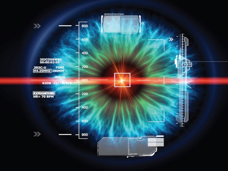
One of the medical fields where the photonic technologies are particularly useful is ophthalmology. They can be used for the diagnosis and treatment of different types of eye disorders and as a tool for surgeries and other medical procedures. A very common example nowadays is the use of lasers for refractive eye surgery. The LASIK, LASEK and PRK techniques are used to correct myopia, hyperopia and astigmatism by removing the cornea or using a cutting laser to precisely shape it. LASIK is probably the most popular technique nowadays and it normally requires two types of lasers for the different processes: a femtosecond laser for cutting – because the ultrafast pulses avoid mechanical and thermal damages in the tissue – and an excimer ultraviolet laser to perform the modification of the cornea, without affecting the other parts of the eye.
Capsulotomy and iridotomy treatments are other good examples of photonics in action. Several millions of cataract surgeries are performed worldwide each year, with an important incidence of post-operative secondary cataracts. Nd:YAG laser capsulotomy has been widely used as an effective treatment for posterior capsule opacification (PCO). It avoids complications such as endophthalmitis and vitreous losses and improves visual acuity in patients after the treatment. Although Nd:YAG laser capsulotomy is accepted as a standard treatment for PCO, it can present some complications. In order to overcome them, several new developments have been made to these lasers. The goal is to enable more efficient, accurate and safe treatments with the most versatile platform possible. As an example, the high-quality optics used in the new Capsulo laser, from Lumibird Medical, allow for better visualisation of the structures in the anterior and posterior segments of the eye. This laser also includes a variable height light tower, which offers dual illumination angles of 16° and 7.5° for improved diagnostics and treatment of both segments.
Diagnostic imaging devices
Medical diagnosis has also benefitted from the advantages of photonic technologies, particularly those based on imaging. In general, we can call medical imaging a set of techniques and processes to get images of the interior of a body for clinical analysis, medical intervention and visual representation of the function of some organs or tissues. Several emission sources have been used to obtain images of the human body, including those emitting light. We can mention, for example, the photoacoustic imaging technique, in which several laser pulses are delivered into a biological tissue where some ultrasonic waves are generated, or the functional near-infrared spectroscopy (fNIRS), an optical brain monitoring technique.

Another of these techniques, the Optical Coherence Tomography (OCT), has revolutionised the clinical practice of ophthalmology. OCT is a high resolution, non-contact advanced optical imaging technique developed for non-invasive cross-sectional imaging of biological systems. OCT can provide 2D and 3D images of the different segments of the eye with micrometre resolution. The structural information is derived from the light backscattered or backreflected at the interfaces between the regions of different optical properties within the object. Besides morphology, OCT reveals and visualises such properties of biological objects as velocity of flow (via Doppler effect), birefringence (via polarisation changes) and tissue extinction. It was initially used to obtain high resolution 3D images of the retina, allowing for the detection of morphological changes in the eye and preventing various eye diseases, including glaucoma and macular degeneration. In recent years, a 3D evaluation of the anterior segment of the eye has also become possible, allowing us to understand the optical quality of the human eye.
There are several versions of OCT, depending on the components used and the configuration of the set-up, but swept-source optical coherence tomography (SS-OCT) has stood out over the rest of the options due its high imaging speed. SS-OCT systems can obtain high-resolution images while reducing the negative effect of patient’s eye movements on scan quality. This performance of SS-OCT is mostly attributable to the laser source characteristics, a wavelength-swept laser. Companies such as CareGlance s.r.l. have been working on new lasers to transform OCT into a real-time, portable technique. They have developed new fast and cost-effective swept sources, granting a 1MHz scan speed together with high coherence and resolution. The high speed is achieved by means of a μs-response time tuneable material, while compactness is granted through an optimised micro-optics cavity assembly.
This need for small and cheap OCTs is definitely a trend in the medical device industry. Low-cost, portable and easy-to-use OCT systems will be essential in facilitating widespread use in point-of-care settings, although they must ensure that they offer the necessary imaging performances.

The HandheldOCT EU Project was born with the goal of developing a new generation of handheld OCT systems in the 1060nm wavelength region for optimum tissue penetration, with size and cost beyond state-of-the- art. Photonic integrated circuit technology allows us to meet all these requirements, and in this particular case, the chip will also include freeform optics developed by the company Nanoscribe GmbH & Co. KG. Other approaches for saving costs and reducing the size include changes in the spectrometer and the scanning optics, new system control design or the use of 3D-printing technology for non-critical components.
Retinal oximetry
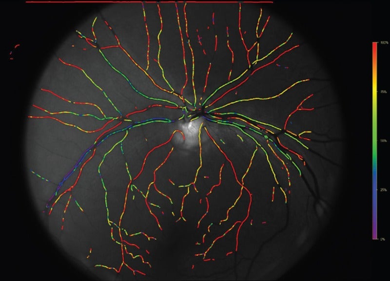
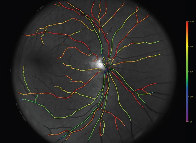
Another interesting diagnostic application of photonics technology is retinal oximetry; that is, the measurement of the haemoglobin in the eye of a patient to assess the amount of oxygen in the bloodstream. Haemoglobin is a protein in the blood that carries oxygen around the bloodstream. It can be used as an indicator of whether a tissue is receiving enough oxygen or not, as any malfunction in the supply of oxygen and nutrients can lead to a variety of diseases. Retinal oximetry is a non-invasive spectrophotometric imaging technique, in which the difference in light absorbance of deoxyhaemoglobin and oxyhaemoglobin is measured. This procedure allows healthcare professionals to detect a lack of oxygen associated with the eyes, such as corneal hypoxia.
“Photonic technologies in ophthalmology and visual sciences are in constant evolution. Several of them have become successful commercial products, and many others are expected to come in the near future.”
Commercial oximeters are based on a stationary fundus camera and they are restricted to patients who can be positioned upright. Some new developments have been performed for supine patients, such as young children, comatose or sedated patients, in cases where measurement of retinal saturation is not possible using current devices. The company Demcon Focal B.V. has developed a handheld, mobile retinal oximeter named Corimap Camera. Apart from the flexibility and mobility, some of its advantages are the use of polarised light (because it could have a beneficial effect on the oxygen measurements due to the reduction of vascular reflections) and the reduction of light exposure with the use of infrared light for the alignment, instead of continuous white light. Some clinical trials were recently published, showing that the Corimap Camera prototype is sensitive to oxygen changes in retinal vessels and performs well in comparison with the current standards of retinal oximetry. The ultimate goal is to use this device on prematurely birthed babies to assess the risk of – as well as act to prevent – eye diseases resulting from oxygen therapy used due to the premature development of their lungs.
Visual simulators
In recent years, we have seen an increase in the number of surgeries to implant intraocular lenses in human beings. Providing patients with the visual experience before the treatment is an important factor in their decision to undergo the operation, so several visual simulators have been developed as clinical instruments to demonstrate the effect of different lens designs and to guide the patient and eye care practitioner in the selection of the most suitable correction. These visual simulators use adaptive optics (AO) to introduce virtually any desired aberration profile and to evaluate the patient’s spatial vision for each controlled aberration profile.
AO visual simulators based on deformable mirrors, spatial light modulators or opto-tuneable lenses are increasingly used to simulate vision through different multifocal lens designs. These active optical elements reproduce the equivalent phase map of a certain optical design in a plane conjugate to the subject’s pupil plane, while the observer is looking at a visual stimulus. The visual simulators were initially limited to experimental environments, given their relatively high complexity and dimensions – although some have made their way into commercial products. One example is the SimVis Gekko from the company 2Eyes Vision, developed for the pre-surgical simulation of presbyopia (age-related loss of focus on nearby objects) correction. Another interesting and high potential application for AO visual simulators is related to the understanding the true impact of aberrations on vision. Once ocular aberrations have been correctly measured, the question arises about their influence on vision. There is still some work to be done in this field.
Photonic technologies in ophthalmology and visual sciences are in constant evolution. Several of them have become successful commercial products, and many others are expected to come in the near future. Certainly, validation and compliance of the regulations will limit their access to some applications, but they can replace several traditional techniques with all their great advantages.


